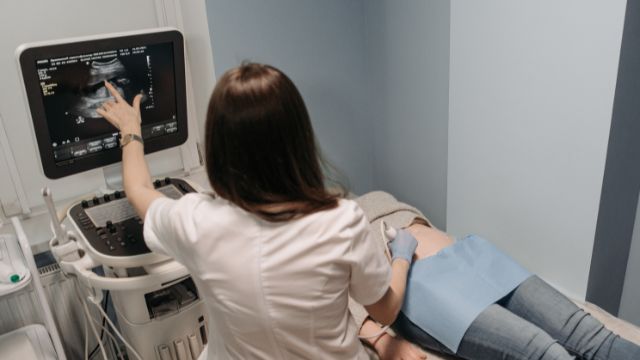
In the last few decades, there has been a growing use of the medical diagnostic procedure called Ultrasound. The process, which is more well known as sonography, happens to be a non-invasive imaging technique. In this process, high frequency sound waves are used for producing images of the body’s internal structure. Here in this short post, we will be offering you a brief regarding the advantages and disadvantages of ultrasound and more information.

Non-invasive ultrasonography stands out in medical imaging. CT and X-rays employ ionising radiation, whereas ultrasound does not. Its unique characteristic makes pregnant women and developing foetuses safer. Safety relies on ultrasounds without ionising radiation. Ionising radiation from various imaging techniques may be dangerous if repeated. High radiation exposure may harm foetal development in pregnant women. The non-invasive nature of ultrasonography increases its safety. It provides detailed inside images by sending high-frequency sound waves into the body and collecting echoes. Radiation is eliminated and obtrusive treatment side effects and suffering are reduced.
The flexibility and importance of ultrasound for healthcare personnel come from its dynamic and real-time imaging. The ability to instantly monitor and evaluate dynamic biological processes offers this talent a medical advantage. In foetal monitoring, real-time ultrasonography is essential. Ultrasound monitors the foetus, mother, and child throughout pregnancy. Real-time data collection and analysis of foetal movement and heart rate is necessary. Doctors can carefully track foetal growth using ultrasound. Live foetal movement and heartbeat monitoring increase diagnosis accuracy and timeliness. Early anomaly discovery permits speedy treatment.
Medical ultrasounds give panoramic images of different anatomical landscapes. It suits organs, blood vessels, muscles, and joints. Ultrasonography's versatility and unique benefits make it useful in many medical domains. Obstetrics requires ultrasound during foetal development. Ultrasound lets pregnant women and doctors examine the unborn heart and movements without surgery. Parents and their unborn child connect emotionally throughout diagnosis. Ultrasound helps cardiologists study heart movements. Ultrasound images the heart's chambers, valves, and blood flow. Cardiology improves by providing better cardiac diagnosis and monitoring.
Ultrasound is a vital medical imaging alternative since it is cheaper than MRI and CT scans. Many healthcare facilities use ultrasound for recurrent imaging because of its cost-effectiveness. Compare ultrasound's cost to other imaging technologies to understand its economic value. Ultrasound is cheaper than MRI and CT. Healthcare companies must handle this cost element to optimise resources without compromising diagnostic efficacy. Ultrasound is preferred by cost-conscious healthcare providers for recurring imaging. Ultrasound's economic feasibility enables for more frequent use without expense increases. In obstetrics, cardiology, and chronic disease treatment, continual monitoring or follow-up is essential.
Mobile ultrasound devices make medical imaging more accessible in many healthcare settings. This makes ultrasound helpful in clinics, emergency departments, and remote places. The mobility of ultrasound instruments makes them a viable healthcare delivery solution. Mobile ultrasound equipment provide urban and rural clinics flexibility. This adaptability allows patients in different locations to get fast, high-quality diagnostic imaging without central infrastructure. Ultrasound's mobility helps emergency departments save time. Ultrasound imaging helps doctors make rapid choices and find treatments. In emergencies, rapid diagnosis is crucial.
Imaging soft tissues like the liver, kidneys, and reproductive systems using ultrasound is a huge diagnostic achievement. This imaging method helps identify tumours, cysts, and inflammatory illnesses by taking exact images of these vital tissues. Soft tissues vary in density and composition, making imaging problematic. Ultrasound becomes the main means for high-resolution images, enabling clinicians to observe these structures' complicated details. Fatty liver disease and tumours may be diagnosed by ultrasound by detecting small liver anomalies.
Ultrasound images soft tissues well but not bone or air. Thick material blocks sound waves, resulting in poor visual quality. To take exact inside photos, high-frequency sound waves are transmitted into the body and echoed back. In bone or air, sound waves hit barriers. Hard and rigid bone reflects and absorbs waves, preventing them from reaching deeper tissues. Ultrasonic imaging in air-filled spaces faces similar issues. In certain places, reflections and refractions hinder sound waves. Areas outside these air-filled zones may not get enough sound wave information for good visuals.
Ultrasound imaging quality relies on operator expertise. The sonographer's skill is essential to ultrasound diagnosis since correct and relevant images need finesse. Operator expertise impacts how these photographs are viewed, creating variability that highlights the necessity for skilled operators. As a dynamic imaging modality, ultrasound demands precise transducer handling and real-time analysis. Operator competence impacts image quality, resolution, and accuracy. Technical expertise, anatomy and pathology knowledge, and patient-specific imaging are needed for this competency. Interpretation causes problems. Operators must identify minor characteristics and irregularities in inner structure images to get insights. Thus, sonographer expertise determines ultrasound image interpretation.
Ultrasound is versatile but lower-resolution than MRI or CT scans. This limitation makes visualising microscopic details harder, which may impact imaging structural anomaly detection. Resolution in medical imaging is the ability to discern near details. Unlike MRI or CT scans, ultrasound may not give adequate information for some diagnoses. This constraint is evident when attempting to observe complex anatomical features or tiny aberrations that may assist diagnosis. In precise clinical situations, visualising minute details is challenging. Microscopic abnormalities or early-stage tumours might decrease ultrasound resolution. MRI or CT scans with higher resolution may be utilised for a more complete review.
Ultrasound struggles with heavy bones and hardened tissues. Ultrasonic waves are blocked by these thick tissues, restricting vision beyond or below them. High-frequency sound waves penetrate tissues to generate echoes and detailed images of inside structures in ultrasound imaging. Thicker things like bones and calcified tissues inhibit this process. Ultrasonic wave reflection and absorption reduce penetration and image quality beneath or beyond these strong obstacles. Imaging past bones or calcified tissues to view deep organs or structures reveals the constraint. These areas may be challenging to diagnose and analyse using medical imaging due to visibility concerns.
Ultrasonic imaging relies on patient cooperation. Gas, obesity, and motion impair ultrasound image quality, making diagnosis difficult. Patient movement during ultrasounds may blur images. Even little patient movements might distort photographs and hide critical information. Patients must minimise undesired movements to achieve clear, diagnostic pictures. Ultrasound imaging is complicated by obesity and fat. Dense fat layers may block ultrasound waves, limiting structural visualisation. Fat may also obscure signals, reducing imaging quality and making anatomical features difficult to discern.
The utility of Ultrasound is already proven, so it is futile to judge it from the aforementioned discussion. In the process of medical analysis, its relevance is still much deep and until some severe change occurs in medical technology, it would probably remain so. However, further modifications and technical corrections might be the need of the day so that the cons of the process can be avoided. Then its utility would further enhance in every possible way.
Ref Links: